Microscopy Helps Feed Mills with Quality Control
- Category:
- Animal Utilization
- Virtual Events
An under-used method for quality assessment in the feed industry is microscopy. This technique is not only fast, but it can also play a critical role in the identification of incoming raw materials and minimize the introduction of foreign materials in the storage and manufacturing process.
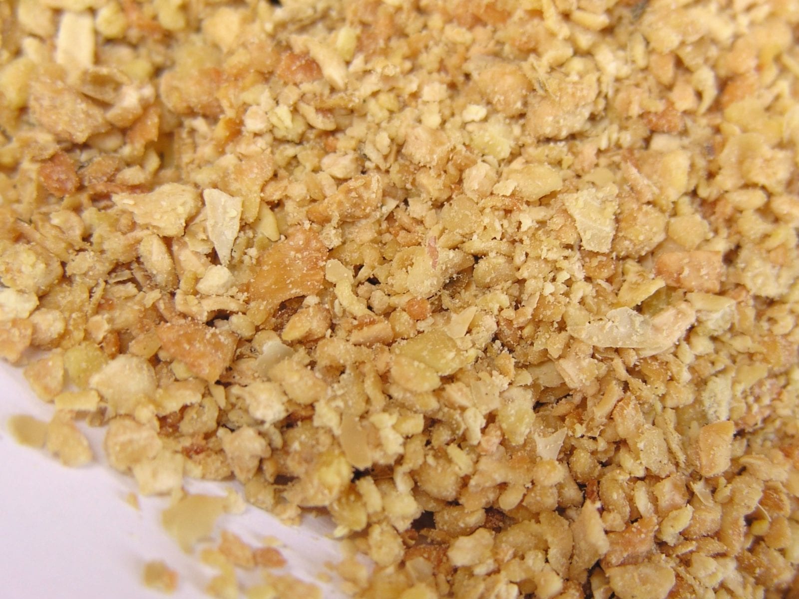
“When ingredients arrive at the feed mill, the quality should be checked before anything is unloaded,” said Dr. Iani Chihaia, a U.S. Soybean Export Council consultant and animal nutritionist in Southeast Europe and Greece. “With a microscope, a lab technician can easily screen for under-toasting, overheating, processing faults, and adulteration of soybean meal.”
He said this is particularly important for soybean meal, which is an expensive ingredient and impacts the quality of compound feeds. This type of exam requires minimal investment in time and resources.
Both, morphological and anatomical structures of the plant do not change. As such, they are easy to identify under the microscope. A trained microscopist should quickly establish the identity and purity of each raw material batch.
“Once the quality is known, the feed mill manager can then decide how to segregate the meal according to quality and origin, and then place appropriately place them,” Chihaia said. “If the exam shows any faults, the lab manager should decide on next steps to maintain the desired quality of the end product.”
While simple, microscopy is an efficient screening method and deserves attention from the quality control managers when evaluating ingredients, especially that of soy.
When looking through the microscope, you should look for uniformity of particle size — one of the quality attributes of U.S. soybean meal, explained Dr. Lene Cecilie Hermansen, a senior microscopy engineer from the Imaging Center of Norwegian Life Sciences University.
Additionally, under the microscope and with different techniques, you can visualize the cell walls, protein bodies, oil cells and the impact of different processing methods, shared Dr. Ninfa Rangel Pederson, a senior scientist in enzymology from the Imaging Center.
This was part of a USSEC live training on feed microscopy, hosted by the European regional office. The two-day training was broadcast from Bucharest, Romania, where more than 250 people participated from more than 50 countries.
During the training, participants learned:
The training included featured speakers Hermansen and Dr. Hilde Raanaas Kolstad, also a senior microscopy engineer from the Imaging Center of Norwegian Life Sciences University, and Rangel Pedersen.